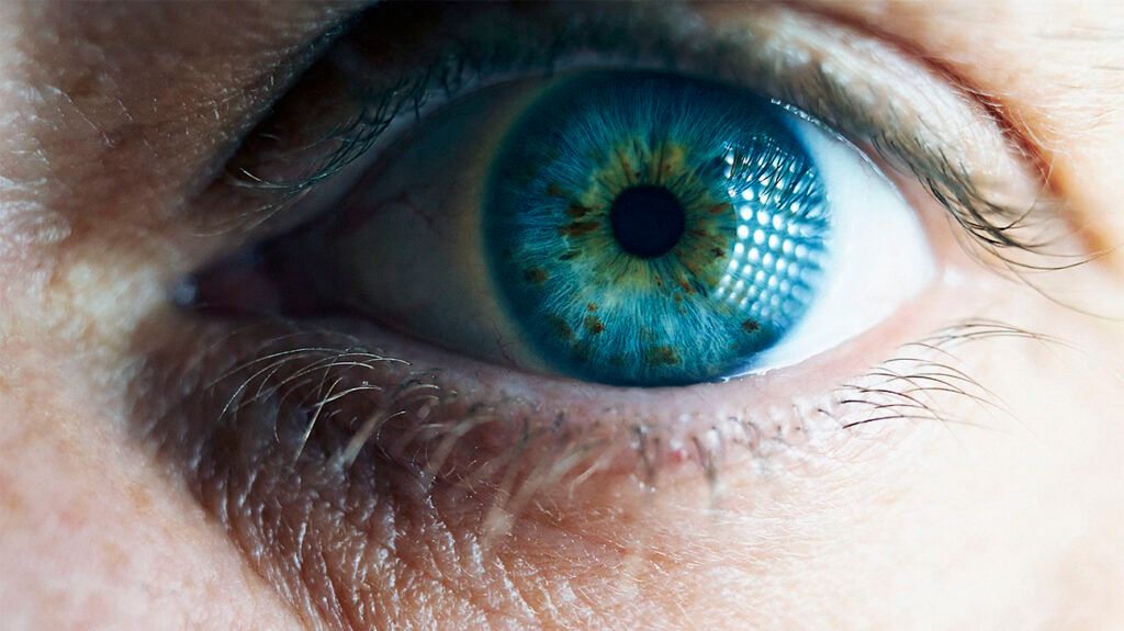
Uveal melanoma (UM) is a rare cancer that develops in the pigment-producing cells within the uveal layer (uvea), which is the middle layer of the eye.
UM is also known as intraocular melanoma.
Early stage UM may not cause any symptoms, and a person may only learn that they have it during a routine eye examination. As the disease progresses, a person may experience symptoms such as a dark spot in the colored part of their eye, a change in the size or shape of their pupil, or changes in their vision.

UM is a
Cancer can develop in any of these parts:
- The iris: This is the colored part of the eye, which gets its color from melanin-producing cells.
- The ciliary body: This is a ring of tissue that sits behind the iris. It consists of muscle fibers that change the shape of the lens so that the eye can focus. It also produces the clear fluid that fills the space between the iris and the cornea (the clear outer layer of the eye).
- The choroid: This is a layer of blood vessels that deliver oxygen and nutrients to the eye. Around 90% of UMs begin in the choroid.
UM of the iris is usually a small, slow-growing tumor that rarely spreads to other parts of the body. In comparison, UM of the ciliary body or choroid is generally larger and more likely to spread to other parts of the body.
Read about the eyes and how they work.
According to the Melanoma Research Alliance (MRA), early stage UM may not cause any symptoms. A person may only learn that they have UM during a routine eye examination with pupil dilation, which is the most effective way to screen for the disease.
As UM progresses, a person may notice the following symptoms:
- a dark spot on their iris
- floaters in their vision
- a change in the size or shape of their pupil
- a bulging eye or a change in the position of the eye in its socket
A person may also experience eye pain or changes in vision, such as:
- blurred or double vision
- reduced vision or loss of vision in one eye
- flashes of light
- floaters
According to the American Academy of Ophthalmology, scientists do not fully understand the cause of UM and other types of ocular melanoma.
However, they know that UM occurs as a result of DNA errors within the melanin-producing cells of the eye. These errors cause the cells to multiply out of control, and the mutated cells then clump together to form a melanoma.
Risk factors for UM
- having fair skin that freckles or burns easily and tans very little or not at all
- having blue, green, or other light-colored eyes
- having a mole, a nevi, or another type of growth in or on the eye
- being over the age of 50 years
- having had excessive exposure to natural and artificial UV light
Importantly, the MRA notes that many people who develop UM do not have any known risk factors for the disease.
When diagnosing UM, doctors will ask about a person’s symptoms and their medical and family history. They will also conduct an initial eye examination.
A doctor may then perform
Dilated pupil eye exam
A dilated pupil eye exam involves using medicated eye drops to dilate the pupil. This allows the doctor to see through the lens and pupil and into the retina and optic nerve.
Ultrasound exam of the eye
An ultrasound exam of the eye involves placing a small probe gently on the surface of the eye. The probe emits high energy sound waves that bounce off the internal structures of the eye and return to the probe as echoes. A computer converts the echo data into a detailed 3D image of the eye’s internal structures.
This test can help determine the location of an intraocular tumor, as well as its size, shape, and thickness. It can also show whether the tumor has spread to nearby tissues.
Transillumination of the globe and iris
Transillumination of the globe and iris involves placing a light on the upper or lower eyelid. The light allows doctors to examine the iris, cornea, lens, and ciliary body.
Eye angiography
Eye angiography is a procedure that allows doctors to investigate blood vessels and blood flow inside the eye.
It involves injecting a special dye into a blood vessel in the arm. The dye then travels around the body and through the blood vessels of the eye. A special camera photographs the retina and choroid to help doctors detect any blood vessel blockages or leakages in these areas.
There are two main types of eye angiography, which use different types of dye: fluorescein angiography and indocyanine green angiography.
Ocular coherence tomography
Ocular coherence tomography is an imaging test that uses light waves to take cross-sectional images of the retina or choroid. This allows doctors to check for swelling or fluid beneath the retina.
After diagnosing UM, a doctor will order further tests to determine whether the cancer cells have spread to other parts of the body. This process is called staging.
According to the
Doctors typically use one of two staging systems to stage eye melanomas.
The American Joint Committee on Cancer (AJCC) TNM system
The AJCC TNM system considers the cancer’s TNM factors, which are:
- T (tumor): the size of the tumor and the extent to which it has grown into nearby structures
- N (nodes): whether the cancer has spread to nearby lymph nodes around the ear or neck or to other parts of the eye
- M (metastasis): whether the cancer has metastasized, or spread to distant parts of the body such as the liver
Doctors will assign a number after each letter to provide additional details about each of these factors. Generally, higher numbers indicate that the cancer is more advanced.
Doctors will then combine a person’s “T,” “N,” and “M” numbers to assign an overall cancer stage. This process is called stage grouping. Uveal melanoma stages range from
The Collaborative Ocular Melanoma Study (COMS) group system
The COMS group system is a simpler staging system that categorizes ocular melanoma into
- Small: The tumor measures between 1 millimeter (mm) and 3 mm in height and between 5 mm and 16 mm across.
- Medium: The tumor measures between 3.1 mm and 8 mm in height and no more than 16 mm across.
- Large: The tumor measures more than 8 mm in height or more than 16 mm across.
Learn more about the stages of cancer.
The treatment for UM depends on several factors, including the tumor size and location and the person’s overall health. Treatment options may include one or more of the following approaches:
- Radiation therapy: Radiation therapy uses high energy particles or waves to destroy or damage cancer cells. It is the most common treatment for UM.
- Surgery: This treatment involves removing the tumor and a small margin of surrounding tissue. Surgery may be an option for small tumors that have not spread beyond the eye.
- Laser therapy: This treatment uses a high energy beam of light to destroy cancer cells. Doctors may use laser therapy to treat small tumors in the front of the eye.
- Chemotherapy: Chemotherapy involves taking chemotherapy drugs, which destroy cancer cells or prevent them from growing. Doctors usually reserve chemotherapy for cases of advanced UM that have spread to other parts of the body.
- Immunotherapy: Immunotherapy uses the body’s immune system to help target and destroy cancer cells. It may be an option for advanced cases of UM. In
2022 , the immunotherapy drug tebentafusp-tebn received FDA approval as a treatment for UM that cannot be removed or has spread to other parts of the body.
The
The SEER database groups these survival rates into three categories according to the degree of cancer spread at the time of diagnosis:
- Localized: There is no sign that the cancer has spread outside the eye.
- Regional: The cancer has spread outside the eye to nearby structures or lymph nodes.
- Distant: The cancer has spread to distant parts of the body, such as the liver.
According to the ACS, the 5-year relative survival rates for UM from 2012 to 2018 were as follows:
- Localized UM: 85%
- Regional UM: 67%
- Distant UM: 16%
How quickly does uveal melanoma spread?
Uveal melanoma may spread at different rates depending on where in the uvea it develops.
UM of the iris is usually a small, slow-growing tumor that
In contrast, UMs of the ciliary body and choroid are generally larger and more likely to spread to other parts of the body.
Is uveal melanoma painful?
In early stages, UM may not cause any symptoms, and a person may not know that they have the disease.
However, as the tumor grows, it may cause eye pain and other symptoms.
Is uveal melanoma hereditary?
Scientists have linked certain genetic mutations to an increased risk of UM.
For example, mutations in the BAP1 gene can cause UM to develop at a young age. These mutations are the result of a
Uveal melanoma (UM) is a rare cancer that develops in the melanin-producing cells in the middle layers of the eye, such as the iris, ciliary body, and choroid. UMs of the iris tend to be less aggressive than those that develop in the ciliary body or choroid. However, UMs of the choroid are much more common.
Early stage UM may not cause any symptoms. As the disease progresses, it may cause symptoms such as a dark spot in the iris, a change in pupil size or shape, and vision changes.
The treatment for UM depends on several factors, including the tumor’s size and location and whether it has spread to other parts of the body. Radiation therapy is the most common treatment option. Other options may include laser therapy, chemotherapy, and immunotherapy.
The outlook for UM depends on the location of the tumor and the stage of the disease at the time of diagnosis. UM of the iris or early stage disease tends to have a more favorable outlook. A routine eye examination can detect early stage UM, so it is important that people receive eye exams regularly.
